26/02/2024
X-ray images of the lung with unprecedented resolution made at Elettra Sincrotrone
Significant improvement in imaging compared with a hospital CAT scan. The study was published in the European Respiratory Journal
Il Parco - Fisica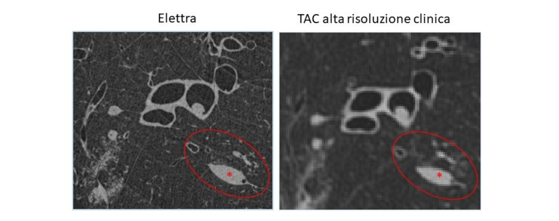
The most important European scientific journal on lung diseases, the European Respiratory Journal, has just published the results of a multidisciplinary research project carried out at Elettra Sincrotrone Trieste, which demonstrated how, thanks to the unique characteristics of the X-rays produced by the Elettra source, it is possible to obtain a significant improvement in X-ray images of the lung compared to a CAT scan made at a hospital. The study, coordinated by Dr. Giuliana Tromba from Elettra, involved the collaboration of Prof. Marco Confalonieri, Director of the Struttura Complessa Pneumologia (Complex Pneumology Structure) of the Cattinara University Hospital in Trieste, and doctors Christian Dullin and Willi Wagner, researchers from the Universities of Göttingen and Heidelberg in Germany.
Currently, with the latest high-resolution CAT machines, it is possible to observe details of the human lung down to the limit of about 0.5 millimeters; however, extending visibility to smaller details would result in a significant increase in the radiation dose administered to the patient. At Elettra, the German-Italian research team demonstrated that, thanks to the innovative ‘phase contrast’ technique, which takes advantage of the peculiar characteristics of synchrotron light (such as monochromaticity and spatial coherence), a view of lung tissue can be obtained with effective resolutions of 0.067 millimeters, i.e., much higher, with a radiation dose to the patient reduced by 2-3 times compared to CAT.
Practically speaking, with this technique it is possible to produce an image of a significantly higher quality by discriminating the various anatomical components and structures much better than with a conventional CAT scan. This makes potentially very useful information available for early recognition of pathological lesions such as tumors and pulmonary fibrosis.
The experiments at Elettra were carried out at the SYRMEP (SYnchrotone Radiation for MEdical Physics) beamline and on an animal model, i.e. pig lungs, which have the property of being the most similar to human lungs. The lungs, obtained as waste from pig slaughtering in Germany, were transferred to Italy and exposed to the Elettra synchrotron light. The newly published results have made it possible to explore the morphology of lung tissue with an unprecedented level of resolution.
With the new Elettra 2.0 light machine currently under construction, which will achieve much higher X-ray energies, these studies can be extended to patients in hospitals in the Friuli Venezia Giulia region. In the examinations of patients, the new technique will allow the densitometric and structural changes caused by different pathologies to be highlighted very effectively, recognising the nature of the pathological lesion more quickly.
According to Prof. Marco Confalonieri “The research work carried out in Trieste will soon allow us to study the human lung as if we had a huge microscope available to us, maximising resolution and mimimising the radiation dose. I am extremely satisfied that our multidisciplinary research work has received this important European recognition. It is the result of the multi-year collaboration between the School of Specialisation in Respiratory Diseases of the University of Trieste and Elettra Sincrotrone Trieste with which we have had an agreement since the opening of the School of Specialisation”.
Dr. Giuliana Tromba is also delighted with the work done and the results obtained, which have been appropriately recognised and enhanced in value by this prestigious publication: “The research team hopes to soon be able to make available to the Trieste healthcare system an innovative way of viewing the alterations created in the lung by serious diseases such as tumors and pulmonary interstitial fibrosis.” The first lung CAT clinical protocol will be aimed primarily at cases where traditional diagnosis leaves interpretive doubts. These are the situations in which the potential of synchrotron light proves crucial in understanding and characterising tissue abnormalities.
The President of Elettra Sincrotrone Trieste, Prof. Alfonso Franciosi, expressed great appreciation for the research carried out, announcing that “from 2026 onwards, when the new Elettra 2.0 light machine becomes operational, a dedicated radiology clinic will be set up on the new SYRMEP-Life Science line which will be able to accommodate patients from all over the Region and will be a unique worldwide resource for medical research”.